The Art of Science
Evident is excited to announce that its 4th Global Image of the Year Scientific Light Microscopy Award is now open for entries! Since its inception, in 2017, the Image of the Year Award has become a celebrated event in the life science microscopy community. We are proud to see that the competition continues to grow each year, with hundreds of entries from across the globe.
At Evident, we are always thrilled to be able to celebrate the beauty and creativity of the life science microscopy community through our Image of the Year Award and we look forward to receiving new and inspiring images that bring out the beauty of science.
From the beginning, it has been our mission to showcase the amazing work of the microscopy community and to help foster a global appreciation for scientific images. We are proud to be part of this celebration that continues to grow each year, and we look forward to seeing the inspiring images that will be submitted this year!
This year, the competition has expanded its scope to include materials science images in addition to life science images. With this, the award seeks to show the world the beauty and versatility of the art of science.
If you have an impressive image you'd like to submit, you can do so by uploading up to three images, along with a description of the equipment used, at Olympus-LifeScience.com/IOTY. Submissions are open until February 28, 2023, so do not wait! Winners will be selected by a jury and announced in the summer of 2023.
Contest details
We are excited to announce the launch of our global science imaging competition!
We have some amazing prizes up for grabs, including:
Global prize: option 1
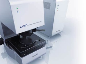
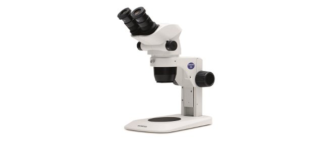
Olympus SZX7 stereo microscope with a DP23 digital camera
Global prize: option 2
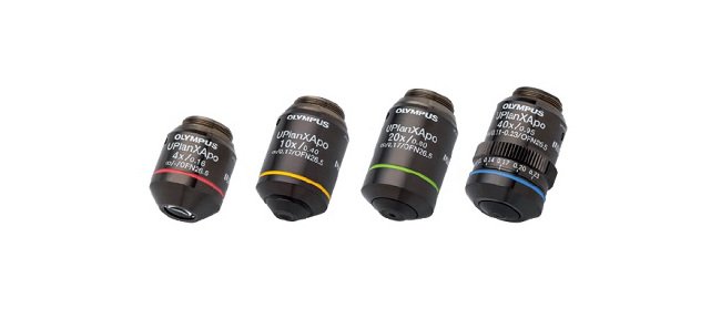
X Line™ objectives for the global winner
Regional prize: option 1
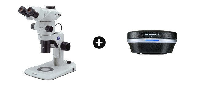
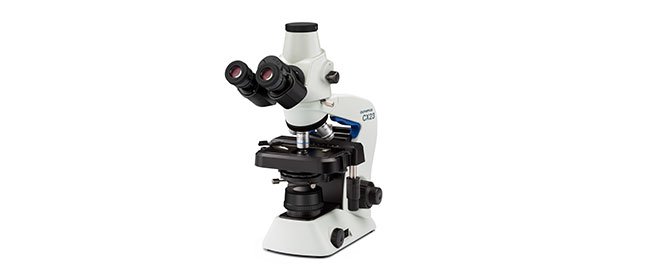
Olympus CX23 upright microscope
Regional Prize: option 2/ Materials Science and Engineering
SZ61 stereo microscope
Plus, a special SZ61 stereo microscope prize will be awarded to the winner of our new dedicated category for materials science and engineering images.
An expert jury of science and imaging professionals from around the world will judge all entries based on their artistic and visual aspects, scientific impact, and microscope proficiency.
Geoff Williams, manager of the Leduc Bioimaging Facility at Brown University; Harini Sreenivasappa, microscopy facility manager of the Cell Imaging Center at Drexel University; Rachid Rezgui, research instrumentation scientist at New York University Abu Dhabi; Yujie Sun, professor and Boya distinguished professor of Peking University; and Sarah Ellis, head of the Centre for Imaging the Tumor Environment (CITE) at the Olivia Newton-John Cancer Research Institute in Australia compose our jury.
So, if you are a promising scientist and have an eye for beautiful images, here is your chance to shine! We cannot wait to see what you produce. Good luck!
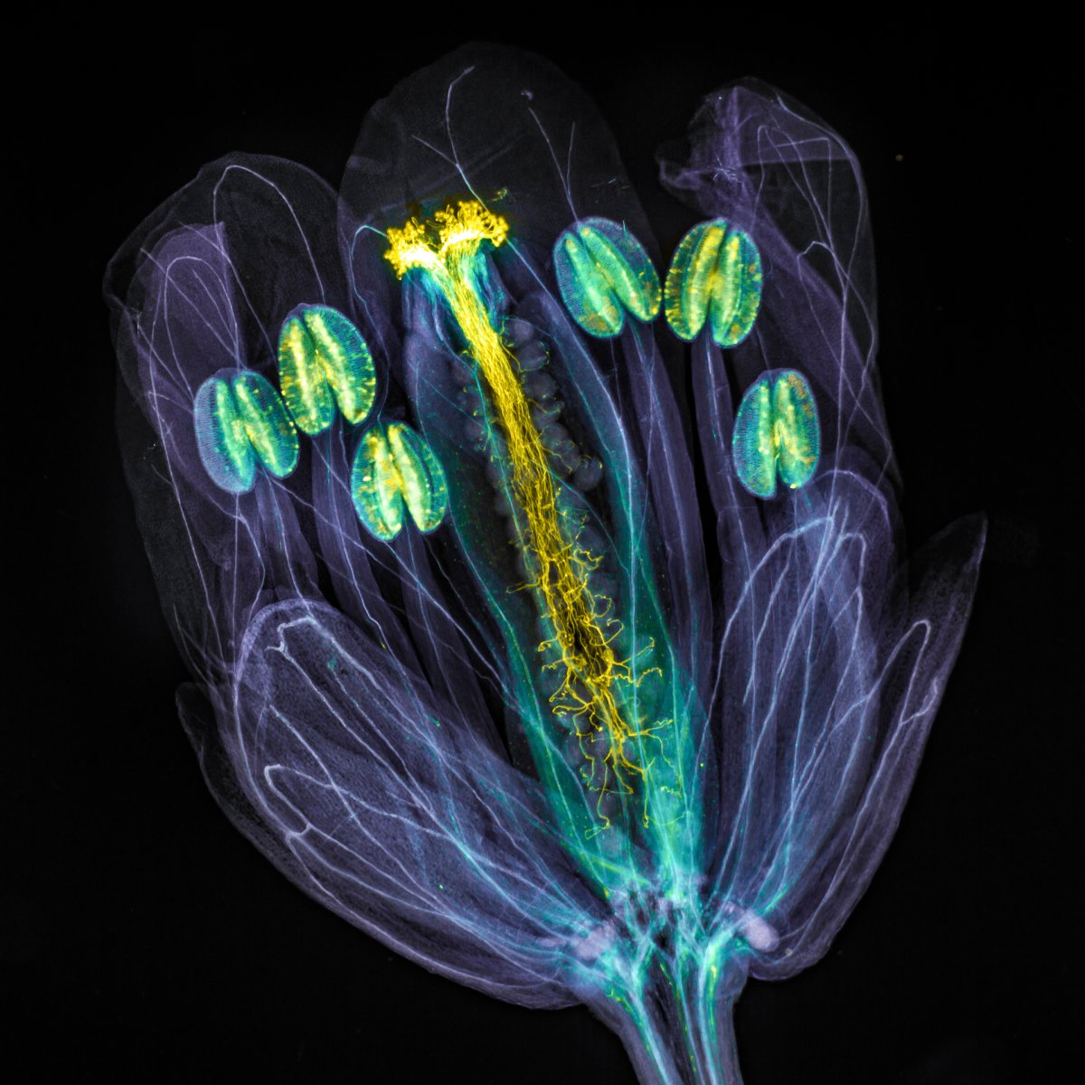
The global winning image was taken by Jan Martinek (Czech Republic).
Arabidopsis thaliana flower with pollen tubes growing through the pistil. The flower tissues were chemically cleared to become transparent, while the pollen tubes were stained with aniline blue (yellow fluorescence) in order to be seen.
Source: Image: